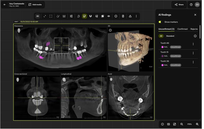
Standfirst: Smart View Detect can flag permanent teeth that may show periapical radiolucency on an existing CBCT scan. The FDA data indicate a meaningful gain in reader sensitivity, but the software is not a general CBCT interpreter, does not justify taking a scan and does not replace a complete clinical review.
Dentsply Sirona has launched Smart View Detect, an artificial-intelligence feature that assists dentists in finding teeth that may be associated with periapical radiolucencies on cone-beam computed tomography scans.
The announcement is significant because dental AI has largely reached practices through two-dimensional bitewing, periapical and panoramic radiographs. Applying computer-aided detection to the much larger three-dimensional CBCT dataset is a more demanding workflow problem.
It is also an easy launch to overstate.
The product described in the US Food and Drug Administration record as DS Core Detect has a deliberately narrow indication. It is a concurrent-reading aid for one type of finding, in permanent teeth, in adults aged 22 or older, using CBCT data that were already acquired for another reason. It is not a replacement for a dentist’s complete review or clinical judgement.
That distinction is more important than the phrase “AI-enabled CBCT diagnosis.”
What the product actually does
Smart View Detect runs inside Dentsply Sirona’s cloud-based DS Core environment. It presents the clinician with a list of teeth that may show periapical radiolucency and a visual map of the suspected area. The dentist can then confirm or reject each flag while reviewing the CBCT.
Dentsply Sirona says the feature became available in the United States and Europe on May 12, 2026. It requires a DS Core Standard or Advanced subscription and is described as compatible with both newly installed and existing Dentsply Sirona CBCT systems.
The operational idea is straightforward: the algorithm performs a second pass across the volume and directs attention to teeth that deserve closer inspection. It does not make the final diagnosis.
| Question | Cleared scope |
|---|---|
| What does it detect? | Permanent teeth that may be associated with periapical radiolucency |
| Which images does it use? | Previously acquired dental CBCT data |
| Who may use it? | Licensed dentists |
| Which patients are included? | Adults aged 22 years or older with permanent teeth |
| Does it recommend taking a CBCT? | No |
| Does it replace the full CBCT review? | No |
| Does it diagnose every CBCT finding? | No |
| Does it make treatment decisions? | No |
The FDA language repeatedly uses “incidental detection.” In practical terms, the CBCT must have been obtained for a separate clinical reason. A positive AI flag cannot retrospectively turn screening for periapical disease into a justification for exposing a patient to CBCT radiation.
What the FDA clearance means
DS Core Detect received Class II 510(k) clearance under reference K253009 in January 2026. The FDA found it substantially equivalent to a legally marketed predicate device, Videa Dental Assist.
Clearance is not the same as an FDA declaration that the software never misses a lesion, that every highlighted area is disease or that the product improves patient outcomes. It means the manufacturer provided evidence supporting substantial equivalence, safety and effectiveness for the stated intended use.
The predicate comparison is noteworthy. Videa Dental Assist analyzes two-dimensional dental radiographs and can detect multiple findings. DS Core Detect analyzes three-dimensional CBCT data but is limited to periapical radiolucency. The regulatory step is therefore important for the imaging modality while remaining narrow in diagnostic scope.
The FDA summary describes two selectable operating points:
- High sensitivity: sensitivity 0.78 and specificity 0.93 in the standalone study.
- Standard sensitivity: sensitivity 0.66 and specificity 0.97.
This is the familiar detection trade-off. A high-sensitivity setting finds more positive teeth but creates more false-positive flags. The standard setting reduces unnecessary flags but misses more positive teeth.
Neither mode is a substitute for systematic review.
The reader study supports assistance, not automation
The most relevant evidence in the clearance summary is a multi-reader, multi-case study involving 11 US readers.
At tooth level, estimated sensitivity increased from 0.421 without assistance to 0.649 with assistance—an absolute improvement of 0.227. Specificity changed from 0.962 to 0.946, a difference of minus 0.016, with the reported confidence interval extending from minus 0.034 to 0.002.
Dentsply Sirona summarizes its internal clinical study as an approximately 46% relative increase in detection without a meaningful increase in false positives. The FDA table is more useful for purchasing and governance discussions because it provides the aided and unaided values directly.
The result supports a role as an attention aid. It does not support autonomous interpretation.
Even with assistance, the study’s tooth-level sensitivity was below 0.65. A negative result therefore cannot be treated as proof that no periapical radiolucency is present. Similarly, a flag indicates a suspicious imaging pattern, not a definitive endodontic diagnosis.
Clinical correlation remains necessary. Symptoms, history, sensibility testing, percussion, periodontal probing, restoration status, prior endodontic treatment and the appearance across multiplanar views all influence what a radiolucency means.
Smaller findings remain harder
The subgroup results show why headline accuracy figures are insufficient.
In high-sensitivity mode, sensitivity was 0.82 for lesions larger than 8 mm³ and 0.65 for lesions at or below 8 mm³. In standard mode, the corresponding values were 0.73 and 0.50.
That gap is clinically relevant. The cases most likely to benefit from an attention aid may include subtle findings that are also more difficult for the algorithm to identify.
Performance also differed around previously treated teeth. In high-sensitivity mode, sensitivity for endodontically treated teeth was 0.82, but specificity was 0.82, compared with sensitivity of 0.72 and specificity of 0.94 for untreated teeth. In standard mode, treated teeth had sensitivity of 0.75 and specificity of 0.90; untreated teeth had sensitivity of 0.56 and specificity of 0.98.
Existing treatment, radiopaque materials, healing change and complex anatomy can affect both human and algorithmic interpretation. Practices should expect a different pattern of flags in an endodontic referral population than in a general dental population.
The evidence outside this product is encouraging but mixed
Recent research supports the general idea that AI can help clinicians identify periapical findings, but the magnitude and direction of the benefit vary.
A 2025 randomized crossover trial involving 30 dentists and 50 panoramic radiographs found that AI assistance increased overall accuracy from 91.6% to 93.3%, mainly by reducing false positives. Sensitivity remained essentially unchanged at about 46%. Junior dentists showed the largest improvements in performance and confidence.
That study involved panoramic radiographs, not Smart View Detect or CBCT interpretation, so it cannot validate this product. It does show that AI’s practical value may come from changing reader behaviour rather than simply producing a high standalone accuracy score.
A separate 2025 study of another commercial AI platform on CBCT scans of molars reported high sensitivity but more moderate specificity in untreated teeth. A June 2026 systematic review of deep-learning systems for periapical radiolucency on panoramic radiographs found promising pooled results but high heterogeneity and notable risks of bias and applicability concerns.
The broader lesson is consistent: performance depends on modality, population, anatomy, treatment status, reference standard and how the dentist interacts with the output.
Workflow and governance questions matter
Because Smart View Detect is delivered through DS Core, the buying decision is partly a software and data-governance decision.
A practice should clarify:
- Which DS Core subscription tier is required in its region?
- Are AI analysis charges included or metered by scan?
- Which CBCT models, acquisition protocols and software versions are supported?
- How long does cloud processing take on the clinic’s actual connection?
- What happens when the internet connection or DS Core service is unavailable?
- Where are images processed and stored?
- Which consent, privacy and data-processing agreements apply?
- Are the AI flags stored in the patient record and audit trail?
- Can a clinician document confirmation or rejection of each finding?
- How will the practice monitor false negatives, false positives and automation bias?
Training should address both over-reliance and dismissal. A clinician may stop searching after seeing no flag, or accept a highlighted area without enough correlation. The safest workflow is to complete a structured review, use the AI output as an additional check and resolve any disagreement deliberately.
Who is likely to benefit
The immediate case is strongest for practices that already use a compatible Dentsply Sirona CBCT system and DS Core. For them, the feature can add a second-reader function without exporting the volume to a separate platform.
General practices that review CBCT only occasionally may value guided attention, although those clinicians also need clear protocols for referral to an oral and maxillofacial radiologist. Endodontic practices could benefit from efficient navigation to suspicious teeth, but the subgroup performance around previously treated teeth deserves particular scrutiny.
Practices outside the Dentsply Sirona ecosystem face a different calculation. A useful AI feature may not justify moving imaging data, subscriptions and review workflows into another platform unless the wider DS Core proposition also makes sense.
The bottom line
Smart View Detect is a meaningful development: an FDA-cleared, cloud-delivered computer-aided detection tool operating directly on CBCT volumes inside a major dental imaging ecosystem.
The strongest evidence is not that the software can read a CBCT by itself. It is that assisted clinicians in the FDA reader study found more teeth with evidence of periapical radiolucency while specificity remained high.
The limitations are equally clear. The indication covers one finding, not the whole volume. It applies to adults with permanent teeth. It is intended for CBCT data already taken for another reason. Smaller lesions were harder to detect. Previously treated teeth showed a different false-positive trade-off. And the dentist remains responsible for the complete review and final judgement.
That is not a disappointing conclusion. It is a realistic description of useful clinical AI: a focused second reader that may reduce misses, provided the practice understands exactly where its responsibility begins and where the algorithm’s clearance ends.
Sources
- Dentsply Sirona, “Dentsply Sirona launches Smart View – Detect,” May 4, 2026. Accessed July 23, 2026.
- US Food and Drug Administration, “510(k) Premarket Notification K253009: DS Core Detect,” January 7, 2026. Accessed July 23, 2026.
- Pul U, Tichy A, Pitchika V, Schwendicke F. “Impact of artificial intelligence assistance on diagnosing periapical radiolucencies: A randomized controlled trial.” Journal of Dentistry. 2025;160:105868.
- Allihaibi M, Koller G, Mannocci F. “Diagnostic accuracy of an artificial intelligence-based platform in detecting periapical radiolucencies on cone-beam computed tomography scans of molars.” Journal of Dentistry. 2025;160:105854.
- Ahmad IA, Ba-Hattab R, Al-Maweri SA, Tamimi F. “Deep learning models for detection of periapical radiolucent lesions on panoramic radiographs: A systematic review and meta-analysis.” International Endodontic Journal. Published online June 25, 2026.
Image credit
Dentsply Sirona, “Smart View – Detect on Canvas,” official press image dated May 2, 2026. Editorial use remains subject to the source’s press-material terms.
Editorial disclosure
This article reports on a manufacturer announcement, an FDA clearance summary and published research. It was independently written, was not sponsored and contains no affiliate links. Digital Dentistry Daily had not independently tested Smart View Detect at the time of writing. Product names and trademarks belong to their respective owners.
About the author